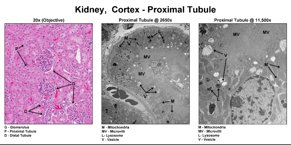
Conventional histopathology uses light microscopy to evaluate stained tissue sections on glass slides, however, some objects are too small to distinguish by light microscopy.
Electron microscopy (EM) is a technique used in biomedical research for obtaining high resolution images to investigate ultrastructural details.


